 MVLscope
A CLOSER LOOK AT NATURE
The butterfly’s secret.
Not just a colour. A structure.
Change the gaps. Watch the wing.

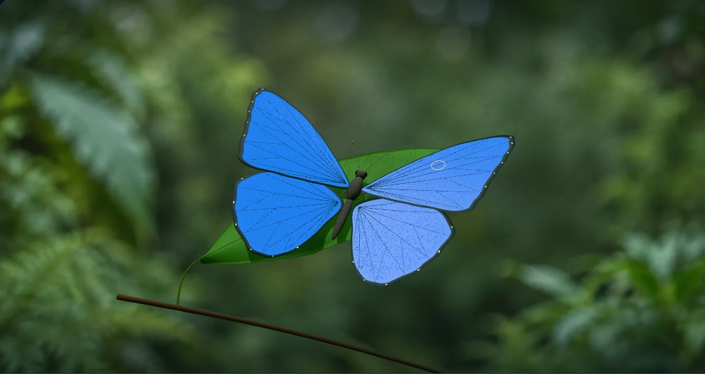
CENTIMETRES → MICROMETRES → NANOMETRES
A wing made of tiny scales.
View
Rotate to inspect. The light-angle control changes colour.
Loading the interactive exhibit · controls will be available shortly
WHY THE COLOUR CHANGES
The gaps pick out the light.
At every boundary, some light reflects. Those reflections can add together, or cancel. Changing the distance changes which wavelengths return strongly.
—strongest visible reflection
in this model Range edge · the true peak may be outside the graph.
Colour uses the whole spectrum, not just its peak.
Your view Baseline at the same angle
An ideal multilayer in air · unpolarized light · approximate display colour
A SMALL EXPERIMENT
Can you bring back the blue?
Start with a 125 nm air gap at 0°. Then increase the light’s angle toward 35° and watch the band move back toward blue. Larger gaps may need angles outside this exhibit’s range.
FROM NATURE TO DESIGN
What would a microscope reveal?
A light microscope can show overlapping scales and, with sufficient resolution and contrast, their ridge lines. Electron microscopy reveals the much finer layered architecture inside the ridges. An electron micrograph itself does not show their optical colour.
Explore how SEM works ↗Inside the model · sources & limits
A structure you can experiment with.
The butterfly and scale geometry are original, Morpho-inspired illustrations. They are not scans of an identified specimen. The wing asset was authored in Higgsfield’s 3D environment; the context still and decorative scale texture are AI-generated illustrations. The soft garden backdrop is also AI-generated. The shaped leaf, branch and contact shadows establish an illustrative natural setting; they are not a reconstruction of a measured habitat. The neutral colour chip and spectrum remain the model output. None of the scenery is measurement evidence.
The editable optical model has seven flat cuticle layers, each 70 nm thick with refractive index 1.56, separated by six air gaps of 80–230 nm. Air surrounds the stack. It calculates s- and p-polarized reflectance using a finite-layer transfer matrix and averages their intensities for unpolarized light. Thicknesses and indices are chosen teaching values. Light and the observer follow a specular arrangement; camera orbit is independent.
The ridge-shaped structure view explains where layers may occur; the spectrum treats each cuticle layer as laterally infinite. Lateral ridge spacing, finite widths, irregularity, curvature, melanin, absorption, wavelength-dependent index and multiple scattering between scales are omitted. Real Morpho wings have a richer and often less angle-dependent appearance. The blue baseline is an illustrative parameter choice, not a fitted spectrum from a particular species. This ideal, lossless stack can reflect almost all light near its strongest band; it should not be read as a measured butterfly’s reflectance. The software recolours the wing as a visual link to the calculation; it is not predicting an entire butterfly’s appearance under arbitrary lighting.
The colour preview integrates the reflected spectrum against CIE D65 daylight data and approximate CIE 1931 two-degree colour-matching functions, then converts to sRGB with out-of-gamut channels clipped to the display range. Monitor gamut, tone mapping and display settings limit the match. Three-dimensional shading adds shape cues; the flat colour chip is the direct spectral preview. The spectrum is sampled every 2 nm. The displayed approximate peak is the strongest sample within 380–780 nm; other maxima may lie outside the visible range.
Scale changes are separate illustrative views, not a continuously calibrated microscope zoom. The nanometre gaps and layer thicknesses in the layer view use the same proportions as the optical model. Colour-control values are physical model inputs; brightness changes are not a scoring system. Nothing here certifies microscopy skills.
Read the research
- Zhang, S. & Chen, Y. (2015) · Nanofabrication and coloration study of artificial Morpho butterfly wings with aligned lamellae layers
- Wang, W. et al. (2013) · Design of a structure with low incident and viewing angle dependence inspired by Morpho butterflies — simulation and design study
- Sosa Espinosa, J., Stavenga, D. G., van der Kooi, C. J. & Giraldo, M. A. (2024) · Morpho butterfly flashiness crucially depends on wing scale curvature
- Kinoshita, S., Yoshioka, S. & Kawagoe, K. (2002) · Mechanisms of structural colour in the Morpho butterfly: cooperation of regularity and irregularity in an iridescent scale
- CIE (2019) · CIE standard illuminant D65 — dataset
- Wyman, C., Sloan, P.-P. & Shirley, P. (2013) · Simple Analytic Approximations to the CIE XYZ Color Matching Functions
- Giraldo, M. A., Yoshioka, S., Liu, C. & Stavenga, D. G. (2016) · Coloration mechanisms and phylogeny of Morpho butterflies
- Nikon MicroscopyU · Resolution — microscopy reference
D65 data: CIE (2019), DOI 10.25039/CIE.DS.hjfjmt59. The 10 nm subset and interpolation are shared under CC BY-SA 4.0. Data attribution and modifications.
These sources support the principles. They do not endorse or validate MVLscope.