01 / UNDERSTAND · 15–20 MIN
Before the Image
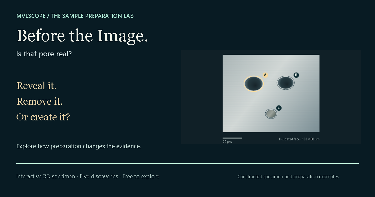
Is that pore real? Explore how sectioning, particle loss and smearing change the evidence in a linked 3D specimen and image.
 Explore the principles →
 MVLscope / Sample preparation
BEFORE THE MICROSCOPE
What you do before imaging shapes what you can learn from it. Start with the principle, then follow a specimen through a virtual preparation workflow.
01 / UNDERSTAND · 15–20 MIN
Is that pore real? Explore how sectioning, particle loss and smearing change the evidence in a linked 3D specimen and image.
Explore the principles →02 / WORK THROUGH IT · 20–30 MIN
From sample to micrograph: select a region, support the specimen, prepare the surface, inspect between stages and choose an examination.
Follow one virtual metal specimen. Compare preparation histories and keep an inspection notebook.
Enter the preparation lab →CHOOSE WHAT YOU NEED TO PRESERVE
Each workbench follows one constructed specimen through decisions that change what you can conclude. Start with the question, keep checkpoints and compare another virtual specimen.
SEM PREPARATION
Prepare one metal or insulating mineral fragment for a surface question or elemental question. Follow the face of interest onto a stub, trace a possible charge path, and record what a coating adds to the evidence.
TEM / FIB PREPARATION
Follow one buried target from a solid into a supported thin lamella. Keep track of what survives, what overlaps, and what preparation may have changed.
BIOLOGICAL / LFM PREPARATION
Follow one synthetic cell culture through an intracellular immunofluorescence workflow. Every committed operation stays in its history; compare a new replicate when a different route is needed.
Choose a workflow for the information you need: a materials cross-section, a surface for SEM, a target in a TEM lamella, or labelled biological structure. Each has distinct limits. No single preparation route suits every specimen.
Choosing a microscopy method →BUILD & DISCOVER
Place an objective, choose a camera and make your first image. Compare how the same specimen changes with your design.
Open Microscope Maker →