
TINY WORLD / COME AND PLAY
There’s a tiny
world in here.
You don’t need to know microscopy.
Just bring a little curiosity.
Free to try · No sign-up · Go at your own pace
MVLscope is a collection of virtual microscopy labs. This little playground is your first peek inside.

YOUR HANDS. YOUR DISCOVERY.
Can you match the mystery view?
Loading your playground…
PICTURE PUZZLE / 1 OF 3
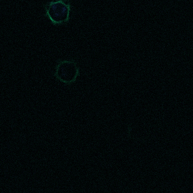
Find this glow.
Match the colours and find a similar focus. Close is enough.
Loading the mystery picture…
01 / FIND A VIEW
A little focus changes a lot.
Move the slider slowly. Which details become sharper? Which fade away?
Look for the blue centres and green strands. This is a model of fluorescently labelled cells.
Use focus and the colour choices below. You can try as often as you like.
About this simulation
These original synthetic cell images come from MVLscope’s LFM teaching model. Focus selects pre-rendered optical sections; colours separate displayed DNA and actin labels. They are not photographs or natural specimen colours. Screen zoom only enlarges the image. This playground changes no lab session and stores no answers.